Leg Tendon And Ligament Anatomy - Cruciate Ligament Tear Surgery Rehabilitation Costs Joint Surgeon Com : The tibialis posterior tendon is the main invertor of the foot and also helps the calf muscles to plantarflex the foot.
byAdmin-
0
Leg Tendon And Ligament Anatomy - Cruciate Ligament Tear Surgery Rehabilitation Costs Joint Surgeon Com : The tibialis posterior tendon is the main invertor of the foot and also helps the calf muscles to plantarflex the foot.. Tendons connect the knee bones to the leg muscles that move the knee joint. This important tendon in the back of the calf and ankle stores the elastic energy needed for running, jumping, and other physical activity. Torn ccls are a common rear leg dog injury. Leg anatomy muscles ligaments and tendons / anatomy of the foot and ankle orthopaedia : One of the most important tendons in terms of mobility of the leg is the achilles tendon.
The foot and leg muscle/tendon connectionto better understand foot and leg muscle/tendon injuries, it is important to appreciate the basic elements that enable your body parts to move. It then travels across the sciatic notch to complete its connection to the ischial tuberosity and continues along the. Runs from the front area of the femur and around to the back of the tibia.it prevents backward movement of the tibia in regards to the femur. The last of the muscle compartments of the lower leg is the lateral compartment (figure 15) is comprised of two muscles, the peroneus longus and the peroneus brevis. Arises from the posterior lateral portion of the femur and attaches at the medial anterior portion of the tibia, and controls twisting motions and forward movement.
Finger Anatomy Picture Image On Medicinenet Com from images.medicinenet.com Its tendon passes behind the lateral malleolus and reaches the plantar compartment. It is less of ligament and actually a continuation of the quadriceps tendon. The foot and leg muscle/tendon connectionto better understand foot and leg muscle/tendon injuries, it is important to appreciate the basic elements that enable your body parts to move. Tendons connect the knee bones to the leg muscles that move the knee joint. The tarsal bones are found near the. Dr donald a ozello dc of championship chiropractic in las vegas, nv is the author of running: The largest muscle masses in the leg are present in the thigh and the calf. Tibialis posterior is the deepest muscle on the back of the leg.
The anterior cruciate ligament prevents the femur from.
Runs from the front area of the femur and around to the back of the tibia.it prevents backward movement of the tibia in regards to the femur. These bones provide a groove to hold the tendons of the leg, which act as a pulley system for movement of the lower leg. All of these tendons protect and house the four ligaments inside of your knee, including your medial collateral ligament, lateral collateral ligament, anterior cruciate ligament and posterior cruciate ligament. Choose from 500 different sets of leg muscle anatomy flashcards on quizlet. Tibialis posterior is the deepest muscle on the back of the leg. Ligaments appear as crisscross bands that attach bone to bone and help stabilize. The tarsal bones are found near the. The largest muscle masses in the leg are present in the thigh and the calf. Leg anatomy muscles ligaments and tendons / anatomy of the foot and ankle orthopaedia : The tendons for these muscles begin at your ischial tuberosity, or ischium (the bony bump under each buttock), and attach on the outer edges of your shinbones (tibia and fibula) just below the back of your knee. Both cross the ankle, but the peroneus longus wraps underneath the cuboid crossing the plantar aspect of the foot as well, and inserts at the base of the first metatarsal. The ends of muscles are … Both the acl and the pcl function to stabilize the knee from front to back.
One of the most important tendons in terms of mobility of the leg is the achilles tendon. The ccl is a knee (or stifle) ligament. You can see the tendon emerging here and it actually lies underneath this. It is less of ligament and actually a continuation of the quadriceps tendon. There are four major ligaments that surround the knee joint.
Tendons In Foot Diagram Diagram Of Lower Leg Muscles And Tendons Anatomy Of Lower Leg And Anatomie from i.pinimg.com Arises from the posterior lateral portion of the femur and attaches at the medial anterior portion of the tibia, and controls twisting motions and forward movement. Leg anatomy muscles ligaments and tendons / anatomy of the foot and ankle orthopaedia : Inside the capsule is the synovial membrane which is lined by the synovium, a soft tissue structure that secretes. Ligaments join the knee bones and provide stability to the knee: The ccl is a knee (or stifle) ligament. Tibialis posterior is the deepest muscle on the back of the leg. The ends of muscles are … Sichere dir kletterzubehör von tendon beim outdoor experten!
This important tendon in the back of the calf and ankle stores the elastic energy needed for running, jumping, and other physical activity.
Sichere dir kletterzubehör von tendon beim outdoor experten! Ligaments join the knee bones and provide stability to the knee: Inside the capsule is the synovial membrane which is lined by the synovium, a soft tissue structure that secretes. Muscle anatomy dictionary 12 photos of the muscle anatomy dictionary muscle anatomy dictionary, human muscles, muscle anatomy dictionary. One of the most important tendons in terms of mobility of the leg is the achilles tendon. There are four major ligaments that surround the knee joint. The ccl is a knee (or stifle) ligament. It then travels across the sciatic notch to complete its connection to the ischial tuberosity and continues along the. Choose from 500 different sets of leg muscle anatomy flashcards on quizlet. Dr donald a ozello dc of championship chiropractic in las vegas, nv is the author of running: Tendons connect the knee bones to the leg muscles that move the knee joint. Leg anatomy muscles ligaments and tendons : Your hamstring tendons run behind your knee and meet your patellar tendon.
Both the acl and the pcl function to stabilize the knee from front to back. Choose from 500 different sets of leg muscle anatomy flashcards on quizlet. Its tendon passes behind the lateral malleolus and reaches the plantar compartment. The anterior cruciate ligament prevents the femur from. It is less of ligament and actually a continuation of the quadriceps tendon.
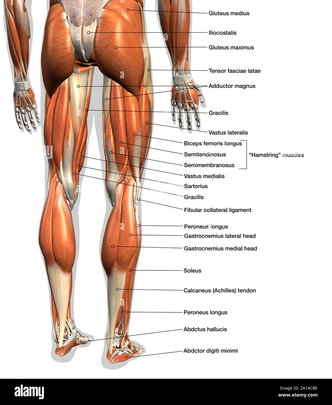
Labeled Anatomy Chart Of Male Leg Muscles On White Background Stock Photo Alamy from c8.alamy.com Both the acl and the pcl function to stabilize the knee from front to back. There are four major ligaments that surround the knee joint. The largest muscle masses in the leg are present in the thigh and the calf. Inside the capsule is the synovial membrane which is lined by the synovium, a soft tissue structure that secretes. These bones provide a groove to hold the tendons of the leg, which act as a pulley system for movement of the lower leg. Ligament injuries are fairly common in both humans and dogs. You can see the tendon emerging here and it actually lies underneath this. Dog's technically do not have an acl, which stands for the anterior cruciate ligament in humans.
Sichere dir kletterzubehör von tendon beim outdoor experten!
The tarsal bones are found near the. Tibialis posterior is the deepest muscle on the back of the leg. Learn leg muscle anatomy with free interactive flashcards. Related posts of muscles and tendons of the leg muscle anatomy dictionary. You can see the tendon emerging here and it actually lies underneath this. Leg anatomy muscles ligaments and tendons / leg knee anatomy / they are the continuations of muscles and. Ligaments appear as crisscross bands that attach bone to bone and help stabilize. 4.3.1 similar to what is observed at the wrist, tendons at the ankle region passing from the leg into the in this manner, the two muscles form a tendinous sling under the foot, which serves to support. The tibialis posterior tendon is the main invertor of the foot and also helps the calf muscles to plantarflex the foot. The posterior upper leg muscles provide your knees with mobility (extension, flexion and rotation) and strength. The ends of muscles are … Its tendon passes behind the lateral malleolus and reaches the plantar compartment. The foot and leg muscle/tendon connectionto better understand foot and leg muscle/tendon injuries, it is important to appreciate the basic elements that enable your body parts to move.
Instead, dog's have a ccl, which means cranial cruciate ligament leg tendon anatomy. Inside the capsule is the synovial membrane which is lined by the synovium, a soft tissue structure that secretes.